
Views: 0 Author: Site Editor Publish Time: 2026-09-15 Origin: Site


Heavily calcified coronary stenoses remain a persistent challenge in interventional cardiology. Calcification serves as a primary predictor of procedural complications and long-term target lesion failure. Standard percutaneous transluminal coronary angioplasty relies on vessel stretching, a mechanism that fails completely in rigid, calcified arteries. Forcing expansion with standard equipment in undilatable coronary stenoses risks severe vessel trauma, flow-limiting dissections, or severe stent underexpansion. Over the past 40 years, clinical approaches evolved from aggressive calcium debulking to modern plaque modification. Specialized balloon designs now serve as the evidence-based bridge between unyielding calcium and optimal stent deployment. Scoring, cutting, and lithotripsy-enabled devices change the treatment paradigm entirely. Selecting the right PTCA Balloon Catheter fundamentally alters lesion compliance, mitigates barotrauma, and ensures procedural success in complex cardiovascular interventions.
Plaque Modification Over Removal: Specialized PTCA balloon catheters do not extract calcium; they create discrete micro-fractures in one or multiple zones of the plaque to fundamentally increase vessel elasticity and compliance.
Targeted Force Application: Advanced designs (scoring, cutting, IVL) allow for effective calcium modification at lower inflation pressures, significantly reducing the risk of barotrauma and severe dissections.
Stent Optimization: Proper lesion preparation with the correct balloon catheter directly correlates with achieving an optimal Minimum Stent Area (MSA), reducing the risk of stent thrombosis and restenosis.
Evidence-Based Selection: Choosing between non-compliant, scoring, or lithotripsy balloons requires intravascular imaging (IVUS/OCT) to assess calcium depth, arc, and thickness.
Calcified plaque exhibits immense mechanical resistance, fundamentally altering the compliance of the coronary artery. Standard compliant balloons are designed to stretch healthy tissue rather than break dense mineral deposits. When an operator inflates a standard balloon inside an undilatable lesion, the fluid dynamics follow the path of least resistance. This creates the classic "dog-bone" effect visible on fluoroscopy. The balloon over-expands in the adjacent healthy vessel segments proximal and distal to the lesion, while completely failing to yield the calcified stricture in the middle.
This physical mismatch leads to catastrophic procedural failures. Applying higher atmospheric pressure only worsens the dog-bone shape. The healthy vessel wall absorbs the excess pressure, stretching beyond its elastic limit. This dynamic frequently results in severe edge dissections, where the intima tears away from the media. Standard devices simply lack the focal force required to fracture rigid mineral deposits. They distribute pressure evenly across their entire surface area, which is highly inefficient when trying to crack a focal plate of calcium.
Unprepared calcium directly causes asymmetric stent expansion. When a stent is deployed into a rigid, unmodified calcified bed, the stent struts fail to appose tightly against the vessel wall. This leaves a mechanical gap. Blood flow becomes turbulent around these malapposed struts, creating areas of low shear stress. Platelets aggregate rapidly in these turbulent zones. Clinical consequences escalate rapidly from this single mechanical failure.
Patients face significantly increased rates of Target Lesion Revascularization (TLR). Stent thrombosis risks multiply when expansion is incomplete, turning a routine intervention into a life-threatening emergency. Myocardial infarction becomes a tangible threat if the vessel occludes acutely. Furthermore, underexpanded stents suffer from altered mechanical stress. The continuous pulsatile force of the heart beating against an unyielding calcium nodule can cause stent fracture. The mechanical integrity of the stent itself fails under continuous stress from unyielding calcium, promoting aggressive neointimal hyperplasia and in-stent restenosis.
Coronary angiography severely underestimates calcium burden. It provides a two-dimensional luminogram that misses up to 40% of severe deep calcification. Optical Coherence Tomography (OCT) or Intravascular Ultrasound (IVUS) is mandatory for complex lesion assessment. These tools diagnose calcium morphology accurately, allowing operators to move from guesswork to precision medicine.
Operators must identify superficial versus deep deposits. They must distinguish concentric arcs from eccentric nodules. OCT provides high-resolution, 10-micron imaging of superficial calcium thickness. Clinical protocols often rely on the "Rule of 5s" for OCT: if the calcium arc exceeds 50% of the vessel circumference (180 degrees), the thickness exceeds 0.5mm, and the length exceeds 5mm, standard balloon angioplasty will fail. IVUS penetrates deeper to reveal total plaque volume and is highly effective in aorto-ostial lesions where contrast clearance limits OCT use. This data dictates the exact device strategy. You cannot select the appropriate PTCA Balloon Catheter without understanding the specific lesion architecture.
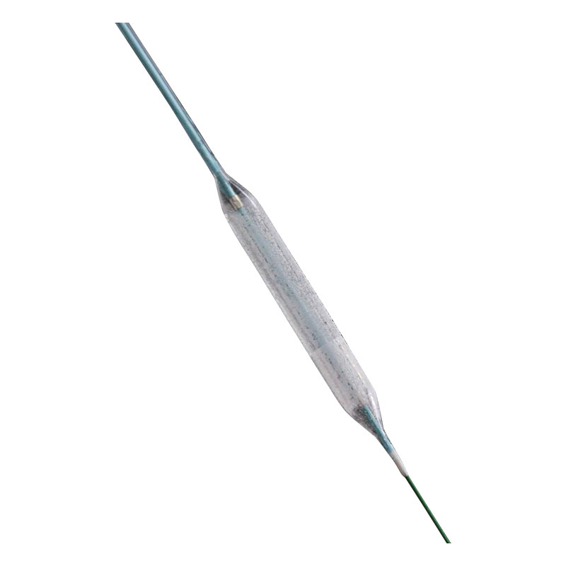
The baseline approach for resistant lesions uses ultra-high burst pressure balloons. Made from non-distensible polymers like PET or Nylon, non-compliant balloons maintain their specified diameter even at extreme atmospheres. They resist the dog-bone effect significantly better than compliant balloons, growing less than 1% in diameter per atmosphere of pressure applied. Operators attempt to force expansion through sheer atmospheric pressure, often inflating these devices to 20 or even 30 atmospheres using specialized inflation devices.
However, severe limitations exist with this brute-force approach. High pressures carry a significant risk of vessel perforation, especially in eccentric lesions. When the balloon pushes against a rigid calcium plate on one side, the force redirects into the healthy tissue on the opposite wall. NC balloons cannot fracture deep or thick calcium arcs effectively. They rely entirely on stretching the vessel wall. When the calcium arc exceeds 180 degrees, NC balloons usually fail to achieve adequate luminal gain. They remain useful primarily for mild to moderate calcification or for post-dilation of deployed stents.
These devices embed atherotomes or scoring wires onto the balloon surface, fundamentally changing how pressure transfers to the vessel wall. These external elements concentrate focal force during inflation. Instead of distributing 10 atmospheres of pressure across the entire balloon surface, the pressure concentrates entirely on the microscopic edges of the blades or wires. They create discrete incisions in the fibrotic and calcified plaque.
The mechanism cracks calcified zones in multiple planes. This ensures controlled fault lines rather than uncontrolled dissections. Cutting balloons utilize three to four micro-blades mounted longitudinally to slice into the plaque. Scoring balloons use nitinol wires wrapped in a helical pattern to score the surface. Both designs prevent the "watermelon seeding" effect, where a standard balloon slips forward or backward out of the lesion during inflation. They modify plaque at much lower pressures (typically 6 to 8 atmospheres) than standard NC balloons. This targeted force application fundamentally alters lesion compliance safely, preparing the bed for optimal stent expansion.
IVL represents a massive leap in calcium modification, integrating ultrasonic transmitters within a low-pressure balloon system. It delivers pulsatile sonic pressure waves directly into the vessel wall. The operator inflates the balloon to a sub-nominal pressure of just 4 atmospheres—enough to ensure the balloon surface apposes the vessel wall without stretching it. The generator then delivers electrical pulses that vaporize fluid within the balloon, creating an expanding and collapsing bubble.
This rapid expansion creates acoustic shockwaves equivalent to 50 atmospheres of localized pressure. These waves crack deep and superficial heavy calcification safely. The system does not rely on high atmospheric pressure stretching the artery. The acoustic waves pass harmlessly through soft, elastic tissue but selectively fracture dense, rigid calcium deposits. This technology proves particularly effective for de novo calcified lesions prior to stent placement. It softens the entire lesion bed without causing severe barotrauma to the healthy vessel wall, making it highly effective for concentric, unyielding calcium cylinders.
Lesion morphology dictates device selection entirely. Superficial calcium responds exceptionally well to cutting and scoring balloons. The blades or wires easily penetrate the surface layer, creating the necessary fault lines. Deep or nodular calcium requires different tactics. Surface modification cannot reach deep deposits, making IVL or adjunctive atherectomy necessary. IVL shockwaves penetrate deep into the medial layer to fracture calcium that blades cannot reach.
Untreated de novo lesions require different strategies than fibrotic in-stent restenosis (ISR). For ISR, scoring balloons excel at preventing balloon slippage and modifying the fibrotic tissue without damaging the existing stent struts. Tortuous anatomy presents another major challenge. Rigid specialty balloons face severe deliverability limitations. Highly trackable standard NC balloons navigate complex bends much better. Operators must constantly balance modification power against device deliverability.
Crossing profiles determine procedural feasibility. Tip diameter and folded balloon profile matter immensely when navigating a 6 French guide catheter through tortuous iliac and coronary anatomy. Standard NC balloons offer excellent crossing profiles, often as low as 0.026 inches. They track easily over standard guidewires.
Advanced modification features add significant bulk. Cutting balloons possess rigid metal atherotomes. This makes them stiff and difficult to deliver through vessels with 90-degree bends or heavy proximal calcification. Scoring balloons offer slightly better flexibility due to the helical wire design. IVL balloons carry integrated emitters and a thicker balloon material, increasing their crossing profile significantly. Operators trade trackability for modification power. Navigating complex, angulated vessels requires careful device selection. A powerful modification tool is useless if it cannot reach the target lesion.
Safety profiles vary drastically across modalities. Flow-limiting dissections and perforations remain primary concerns during complex interventions. Ultra-high-pressure NC balloon inflation maximizes vessel trauma risks. The sheer force required often tears adjacent healthy tissue, leading to spiral dissections that require multiple bailout stents.
Low-pressure dilatation mitigates this barotrauma significantly. IVL operates at very low atmospheres, relying on acoustic energy rather than mechanical stretching. Scoring balloons also operate at lower pressures due to focal force concentration. Minimizing barotrauma prevents edge dissections. It ensures a stable, healthy landing zone for the subsequent stent deployment, reducing the overall length of metal required to fix the vessel.
Technology | Primary Indication | Mechanism of Action | Crossing Profile | Barotrauma Risk | Optimal Calcium Type |
|---|---|---|---|---|---|
Non-Compliant (NC) | Mild/Moderate Resistance | High-pressure stretching (up to 30 atm) | Excellent (Highly trackable) | High at burst pressures | Mild, non-circumferential |
Scoring/Cutting | Superficial/Fibrocalcific | Focal force incision via blades/wires | Moderate (Stiffer profile) | Medium | Superficial, eccentric |
Intravascular Lithotripsy (IVL) | Severe/Deep/Concentric | Sonic pressure waves at 4 atm | Bulky (Requires larger guide support) | Low | Deep, concentric, thick |
Standard NC balloons cost significantly less upfront, making them the default choice for routine cases. Premium scoring or IVL catheters carry higher unit economics, often costing ten times as much as a standard balloon. Cath lab directors must evaluate the return on investment carefully, looking beyond the initial purchase price.
Prevented complications save immense hospital resources. A failed procedure using cheap equipment ultimately costs more. Treating a severe dissection or perforation requires expensive covered stents, prolonged fluoroscopy time, increased contrast use, and potentially emergency surgical backup. Reducing multiple balloon exchanges streamlines the entire procedure, freeing up the lab for the next case. Long-term avoidance of Target Lesion Revascularization justifies the initial expense. Investing in proper lesion preparation tools reduces overall healthcare expenditures over the patient's lifetime by preventing readmissions.
Specialized consoles require setup time. IVL generators introduce a slight learning curve for the staff, requiring cable connections and specific fluid preparation to avoid micro-bubbles in the balloon. Standalone balloon catheters deploy instantly straight from the package. Operators must consider the impact on overall fluoroscopy time and radiation exposure.
Complex setups may increase initial procedural duration by a few minutes. However, effective lesion preparation speeds up the stenting phase dramatically. Contrast volume increases if multiple standard devices fail to cross the lesion or fail to expand, requiring repeated angiograms to check progress. Rapid setup protocols mitigate these delays. Well-trained cath lab teams integrate advanced consoles seamlessly. The time invested in proper preparation always pays off during final stent optimization, turning a frustrating two-hour case into a smooth, predictable procedure.
Aggressive lesion preparation carries inherent risks, regardless of the technology used. Operators must establish strict clinical protocols for managing adverse events.
Balloon Entrapment: If a scoring balloon becomes stuck in a heavily calcified lesion, operators must avoid pulling aggressively. Techniques include deep seating the guide catheter or using a buddy wire to create space.
Balloon Rupture: High-pressure inflations can cause pinhole leaks or longitudinal tears. Pinhole leaks create high-pressure jets that can dissect the vessel wall. Always maintain distal wire position. If a balloon ruptures, deflate completely and apply negative pressure before careful withdrawal.
Distal Embolization: Fracturing heavy calcium can release micro-debris, clogging the distal capillary bed and causing the no-reflow phenomenon. Operators must have vasodilators like adenosine or sodium nitroprusside drawn up and ready for immediate intracoronary injection.
Vessel Perforation: Keep covered stents immediately available in the room. If perforation occurs, inflate a standard balloon proximal to the leak to stop bleeding while the team preps the covered stent.
Symmetrical stent apposition defines procedural success. Operators target an MSA greater than 80% relative to the average reference vessel area. Proper plaque modification makes this achievable. Intravascular imaging confirms the final MSA, ensuring no mechanical gaps remain.
Underexpanded stents leave a residual bottleneck. This bottleneck alters hemodynamics negatively, increasing shear stress on the stent struts. Achieving optimal MSA requires the underlying calcium to yield completely. The stent struts must embed slightly into the modified plaque, creating a smooth, circular lumen. A properly expanded stent minimizes the risk of future adverse events. When the PTCA Balloon Catheter fractures the calcium effectively, the stent deploys at nominal pressure, achieving full expansion without requiring dangerous high-pressure post-dilation.
Clinical trial benchmarks validate these modern approaches. Target vessel failure rates drop significantly when calcium is modified properly prior to stenting. Major adverse cardiovascular events decrease across the board in complex patient populations.
Utilizing specialized devices for calcified lesions improves long-term patient survival. Restenosis rates plummet when stents are fully expanded and well-apposed. Late stent thrombosis, a catastrophic event that can occur months or years after the procedure, becomes a rare occurrence. The initial investment in advanced lesion preparation translates directly into durable clinical results. Patients experience less angina, require fewer repeat procedures, and maintain better overall ventricular function.
Audit your current lesion preparation algorithms immediately to ensure alignment with modern plaque modification standards.
Mandate intravascular imaging (OCT or IVUS) for all suspected calcified lesions before finalizing device selection.
Evaluate your specialty balloon catheter inventory against recent clinical guidelines to ensure you have scoring, cutting, and IVL options readily available.
Train cath lab staff on rapid setup protocols for advanced modification consoles to minimize procedural delays and contrast use.
A: Compliant balloons stretch with pressure, risking over-expansion in healthy tissue. Non-compliant balloons maintain their specified diameter even at high pressures. They focus force directly on the stricture without yielding, preventing the dog-bone effect.
A: Cutting balloons feature micro-blades called atherotomes for precise incisions. Scoring balloons use external wires to score the plaque. Scoring devices generally offer a slightly better crossing profile and more flexibility.
A: No. Modern balloon-based techniques do not remove calcium. They fracture and modify the plaque in multiple zones. This increases vessel elasticity, allowing for proper stent expansion without extracting the mineral deposits.
A: IVL is indicated for severe, deep, or concentric de novo calcification. High-pressure balloons pose a severe risk of vessel rupture here. They are also unable to fracture thick calcium arcs effectively.
A: Risks include balloon rupture, vessel perforation, and flow-limiting dissection. Operators may also experience difficulty crossing highly tortuous or tight lesions due to the larger device profiles.
A: Inadequate preparation leads to stent underexpansion. This is the leading mechanical cause of in-stent restenosis. It also significantly increases the risk of late stent thrombosis and target lesion failure.


