
Views: 0 Author: Site Editor Publish Time: 2026-09-15 Origin: Site


The transition in interventional oncology toward minimally invasive procedures changes standard-of-care protocols for localized and metastatic tumors. Oncology departments and hospital procurement committees face a specific operational problem. They must balance capital expenditures with the clinical mandate to treat surgically ineligible patients. Hospitals need to reduce complication rates and optimize bed turnover without compromising oncological outcomes. Integrating a modern Ablation Device offers a scientifically validated, targeted method for tumor necrosis. This guide evaluates the clinical scope, technical evaluation criteria, and operational return on investment of adopting microwave ablation technology in a hospital setting. We will examine how this equipment expands treatment viability, improves thermal dynamics, and drives departmental efficiency.
Expanded Treatment Viability: A microwave ablation device allows hospitals to treat surgically inoperable tumors across hepatic, pulmonary, renal, colorectal, and thyroid indications, particularly in hard-to-reach anatomical locations.
Superior Thermal Dynamics: Compared to legacy radiofrequency systems, microwave ablation provides faster heating, larger ablation zones, and less susceptibility to the heat-sink effect from nearby blood vessels.
Operational Efficiency & Patient Satisfaction: Minimally invasive ablation reduces reliance on intensive care units (ICUs), shortens patient length of stay, accelerates departmental throughput, and significantly improves the patient experience by offering a low-risk alternative to open surgery.
Strategic Procurement: Evaluating an ablation device requires strict scrutiny of antenna design, cooling mechanisms, imaging compatibility, and vendor training support to mitigate implementation risks.
A microwave Ablation Device operates on the principles of dielectric heating. The generator emits electromagnetic waves, typically at frequencies of 915 MHz or 2.45 GHz. These waves travel through a specialized coaxial antenna inserted directly into the target lesion under imaging guidance. The rapidly alternating electromagnetic field forces polar water molecules within the tissue to oscillate continuously. This rapid oscillation generates immense kinetic energy and localized friction. The resulting thermal coagulation induces immediate, irreversible cellular necrosis at temperatures exceeding 60 degrees Celsius.
Unlike traditional modalities, this process does not require an electrical current to flow through the patient's body. Grounding pads are entirely unnecessary. This fundamental difference eliminates the risk of alternate site skin burns. It also allows the system to penetrate tissues with high impedance. When tissue chars and desiccates during heating, electrical conductivity drops. Because microwave energy relies on electromagnetic radiation rather than electrical conduction, it actively pushes through charred tissue, aerated lung, or dense bone to maintain a growing ablation zone.
Interventional radiologists frequently evaluate microwave ablation against radiofrequency ablation (RFA) and cryoablation. Each modality presents distinct technical trade-offs in the clinical setting. Microwave technology consistently achieves higher peak temperatures and faster ablation times than RFA. RFA relies on electrical conductivity, which drops significantly as tissue desiccates and chars during the heating process. This desiccation limits the active heating zone, forcing the operator to perform overlapping ablations for larger tumors. Microwave energy actively radiates through charred and desiccated tissue, creating a more uniform zone of necrosis.
The heat-sink effect represents a major limitation for RFA. When a tumor sits adjacent to a major blood vessel, such as the portal vein, flowing blood cools the surrounding tissue. This convective cooling prevents RFA from reaching lethal temperatures at the tumor margins, leading to local recurrence. A microwave Ablation Device overcomes this obstacle. It generates heat so rapidly and intensely that the cooling effect of nearby vascular structures becomes negligible. This capability maintains therapeutic efficacy near major vessels where RFA typically fails.
Feature | Microwave Ablation (MWA) | Radiofrequency Ablation (RFA) | Cryoablation |
|---|---|---|---|
Energy Source | Electromagnetic waves (Dielectric heating) | Electrical current (Resistive heating) | Argon/Helium gas (Freezing/Thawing) |
Heat-Sink Effect | Minimal susceptibility | High susceptibility | Moderate susceptibility |
Ablation Speed | Very Fast (typically 5-10 minutes) | Moderate (typically 12-30 minutes) | Slow (multiple freeze-thaw cycles) |
Tissue Impedance Limit | No limitation (penetrates char/bone) | Limited by tissue charring/desiccation | Not applicable |
Multidisciplinary tumor boards increasingly incorporate microwave ablation into standard treatment algorithms. Clinical guidelines recognize it as a primary, adjunctive, or palliative treatment modality. For early-stage hepatocellular carcinoma, ablation often serves as a curative primary therapy equivalent to surgical resection for tumors under 3 centimeters. Surgeons also use it adjunctively during open or laparoscopic resections to clear distant hepatic margins, preserving functional liver volume. In palliative scenarios, ablation reduces tumor burden and alleviates localized pain.
Standardizing this care requires robust institutional protocols. Hospitals must align their interventional radiology, surgical oncology, and medical oncology departments. We recommend implementing a standardized workflow to ensure consistent outcomes:
Establish strict patient selection criteria based on tumor size, location, and patient comorbidities.
Mandate pre-procedural cross-sectional imaging (CT or MRI) within 30 days of the planned intervention to map the exact tumor volume.
Standardize intraoperative imaging guidance protocols, specifying when to use ultrasound versus CT fluoroscopy.
Implement a uniform post-ablation imaging schedule to monitor for local recurrence at 1, 3, 6, and 12 months.
Treating hepatocellular carcinoma (HCC) and colorectal cancer liver metastases remains the primary and most validated use case. Many patients present with tumors that are difficult to resect surgically. Factors precluding surgery include large tumor size, multifocal disease, or complex anatomical locations near the hepatic hilum. Poor underlying liver function, such as severe cirrhosis, also disqualifies many candidates from major hepatectomy.
Microwave ablation provides a highly effective alternative. The primary success criterion is achieving complete ablation margins. Operators aim for a 5mm to 10mm margin of healthy tissue surrounding the visible tumor. High-powered microwave systems create large, spherical zones that easily encompass these margins in a single insertion. This capability significantly reduces local recurrence rates in hepatic applications compared to older thermal technologies. For patients with multiple small metastases, operators can use multiple probes simultaneously to ablate several lesions in a single session, drastically reducing the overall treatment timeline.
Thoracic oncologists frequently utilize thermal ablation for primary non-small cell lung cancer (NSCLC). It also serves patients with metastatic pulmonary lesions originating from colorectal or renal primary sites. Many of these patients are medically inoperable due to poor pulmonary reserve, emphysema, or advanced age. A microwave Ablation Device delivers highly targeted thermal energy directly into the lung parenchyma.
This precision preserves healthy lung tissue far better than a surgical lobectomy or wedge resection. It also provides a localized alternative to stereotactic body radiation therapy (SBRT). Interventionalists can safely access hard-to-reach places using CT fluoroscopy guidance. The rapid heating cycle minimizes the time the rigid probe remains in the moving lung. This speed substantially reduces the risk of intraprocedural pneumothorax, bronchopleural fistulas, and other pleural space complications. If a pneumothorax does occur, the short procedure time allows the operator to manage it immediately with a small chest tube before the patient leaves the suite.
Clinical applications continue to expand rapidly beyond the liver and lungs, providing hospitals with broader utility for their equipment. Expanding the case mix ensures the generator is utilized across multiple departments, maximizing the return on the initial capital investment.
Renal Cell Carcinoma: Urologists employ the device for nephron-sparing treatment of small renal masses (typically under 4 centimeters). This approach preserves crucial kidney function in patients with pre-existing chronic kidney disease or solitary kidneys.
Thyroid Nodules: Endocrinologists apply microwave energy to benign and malignant thyroid lesions. Successful ablation shrinks symptomatic nodules, relieves swallowing difficulties, and may help patients avoid lifelong hormone replacement therapy associated with total thyroidectomy.
Breast and Bone: Emerging data supports the use of ablation for comprehensive palliative care. It provides excellent localized control and rapid pain relief for osteolytic bone metastases. Researchers are also actively evaluating its efficacy for specific, early-stage breast cancer profiles in patients refusing surgical lumpectomy.
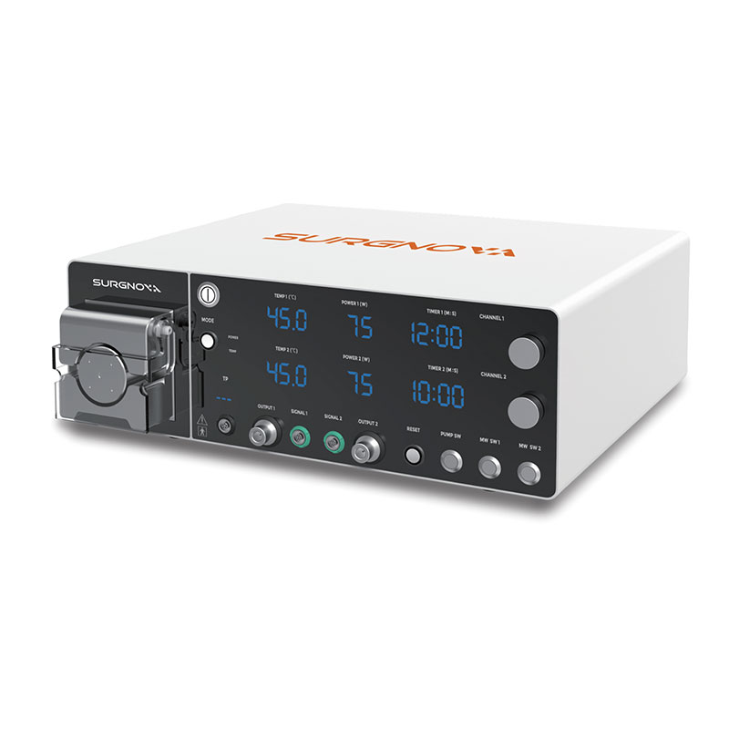
Procurement teams must define strict technical evaluation standards before acquiring new capital equipment. The primary metric is the ability of the generator to deliver consistent, predictable spherical or elliptical ablation zones. Irregular or teardrop-shaped zones increase the risk of leaving residual viable tumor cells at the margins. Operators need reliable wattage control to tailor the energy output to the specific tissue type.
Frequency specifications also play a critical role in clinical outcomes. Systems operating at 915 MHz generally offer deeper tissue penetration, making them suitable for very large tumors. Systems at 2.45 GHz provide faster, more intense localized heating, which is ideal for precise, rapid ablations. Hospitals should evaluate which frequency aligns best with their anticipated departmental case mix. Predictability directly correlates with clinical success and reduced patient readmission rates. When the thermal zone matches the manufacturer's reference charts, the interventional radiologist can plan the probe trajectory with absolute confidence.
The physical design of the probe or antenna dictates procedural safety and maneuverability. Shaft diameter, measured in gauge, impacts the insertion trauma. Thinner antennas (e.g., 14-gauge to 17-gauge) reduce the risk of bleeding, especially in highly vascularized organs like the liver or spleen. Tip design influences how smoothly the probe penetrates dense, fibrotic tumor tissue without deflecting off course. Track-ablation capabilities are absolutely essential. As the operator withdraws the probe, the system briefly emits energy to cauterize the needle tract. This feature prevents malignant tumor seeding along the insertion path and minimizes post-procedural bleeding.
Active cooling mechanisms are mandatory for modern, high-powered systems. Generators circulate sterile water or gas through the antenna shaft during energy delivery. This active cooling prevents the shaft from overheating, thereby avoiding collateral thermal damage to non-targeted soft tissue along the insertion path. It prevents severe, full-thickness skin burns at the percutaneous insertion site. Evaluating the efficiency and reliability of these cooling systems is a critical procurement step for patient safety. Systems that use internal closed-loop fluid cooling generally offer superior thermal regulation compared to older gas-cooled models.
An Ablation Device must integrate seamlessly with existing hospital imaging infrastructure. Compatibility with ultrasound, CT, and MRI guidance systems is non-negotiable. The antenna must be highly echogenic under ultrasound to ensure clear visibility. It must also produce minimal beam-hardening artifact on CT scans to allow accurate assessment of the ablation zone.
Advanced software integrations further enhance targeting accuracy. Modern systems offer 3D mapping and multi-planar trajectory planning. Real-time thermal monitoring overlays assist the interventional radiologist during the procedure. These digital tools help operators visualize the expanding heat zone dynamically. They ensure the lethal isotherm fully encompasses the tumor margin while strictly sparing adjacent critical structures like the gallbladder or central airways. To ensure proper integration, clinical teams should follow a standardized setup sequence:
Verify the generator's software version is compatible with the hospital's PACS and imaging modalities.
Perform a dry run of the trajectory planning software using historical patient DICOM data.
Test the echogenicity of the specific probe models under the exact ultrasound machines used in the interventional suite.
Confirm that the generator cart fits physically within the spatial constraints of the CT fluoroscopy room.
Hospital administrators must contextualize this investment against broader healthcare economic trends. The global tumor ablation market demonstrates robust, sustained growth. Analysts valued the market at USD 1.70 billion in 2024. Projections indicate it will reach USD 4.59 billion by 2032, representing a 13.3% compound annual growth rate. This trajectory reflects a massive, patient-driven shift in demand. Patients actively seek minimally invasive alternatives to traditional open surgeries to minimize recovery time and scarring.
Framing the ablation system as a strategic asset is essential for hospital leadership. It captures this transitioning patient volume and prevents out-migration to competing facilities. It also attracts regional referrals for complex, inoperable cases that smaller community hospitals lack the technology to manage. By establishing a comprehensive interventional oncology program, a hospital positions itself as a regional center of excellence, driving downstream revenue through associated imaging, lab work, and follow-up consultations.
Calculating the operational return on investment requires a comprehensive view of hospital resources and workflows. Administrators must compare the capital cost of the generator and single-use consumables against systemic downstream savings. Ablation procedures significantly reduce operating room (OR) time compared to open resections. They require lower anesthesia levels, often utilizing moderate conscious sedation instead of general anesthesia. This shift minimizes postoperative intensive care unit (ICU) admissions and reduces the burden on anesthesia staff.
The transition toward outpatient or short-stay procedures transforms departmental throughput. Patients typically discharge within 24 hours of a percutaneous ablation. This rapid turnover frees up high-acuity hospital beds for more critical, higher-margin surgical cases. It also improves overall patient satisfaction through low-risk, comfortable treatment pathways. The financial model shifts favorably from high-cost, long-stay surgical DRGs to high-throughput, highly efficient interventional radiology procedures. A single interventional suite can often process three to four ablation cases in the time it takes to perform one complex open hepatectomy.
Financial viability depends entirely on accurate coding and consistent payer reimbursement. Hospitals must navigate current CPT coding and DRG reimbursement structures carefully. Medicare and private commercial payers provide established, mature codes for microwave ablation procedures across various organ systems. Liver, lung, and renal ablations generally feature well-defined, favorable coverage policies.
Documentation requirements are notably stringent. Physicians must clearly document the medical necessity, exact tumor size, and precise anatomical location in the operative note. They must also record the specific imaging guidance modalities used during the procedure. Failure to provide granular, specific documentation inevitably leads to claim denials and delayed revenue. Hospitals should train their billing departments alongside their clinical staff prior to implementation. This dual training ensures the facility captures the full financial value of every procedure performed and minimizes time spent in the appeals process.
User inexperience poses a significant clinical threat during early adoption. Suboptimal ablation margins lead to rapid local tumor recurrence. Misjudging the thermal sphere can cause severe collateral tissue damage to adjacent organs, leading to fistulas or strictures. Hospitals must mandate rigorous, vendor-supplied training programs. Interventional radiologists and surgical oncologists should complete ex-vivo phantom modeling exercises to understand thermal dynamics. Initial clinical cases require direct proctoring by experienced, high-volume users. Establishing a formal internal credentialing pathway ensures baseline competency before independent operation begins.
Procedure delays severely impact departmental schedules and patient trust. Generator downtime or backorders on specific probe sizes force last-minute cancellations. This unreliability damages the department's reputation and disrupts continuous patient care. Procurement teams must evaluate vendor service-level agreements (SLAs) carefully. Strict warranty terms, preventative maintenance schedules, and guaranteed response times for repairs are essential. Hospitals should also negotiate the cost and guaranteed availability of single-use ablation antennas. Maintaining an adequate par level of various probe lengths and gauges prevents supply chain bottlenecks.
Applying the technology to inappropriate tumor profiles yields poor oncological outcomes. Treating excessively large tumors often results in incomplete necrosis. Ablating lesions too close to critical structures causes severe, sometimes fatal, complications. Multidisciplinary tumor boards must establish strict institutional guidelines. These protocols define absolute tumor size limits for single-probe and multi-probe ablation. They mandate safe distance margins from critical structures like central bile ducts, the heart, or the bowel. Guidelines must also address patient coagulation status to prevent catastrophic intraprocedural bleeding.
Request peer-reviewed outcome data and clinical trial results from prospective vendors to validate their specific generator and probe configurations.
Schedule hands-on clinical demonstrations with your interventional radiology team to evaluate probe ergonomics, software usability, and imaging compatibility.
Conduct a facility-specific cost-benefit analysis to quantify the projected impact on bed turnover, operating room utilization, and departmental throughput.
Establish a multidisciplinary committee to draft institutional guidelines for patient selection, intraoperative workflows, and post-procedure monitoring.
A: Microwave devices use electromagnetic fields to oscillate water molecules, generating rapid, localized friction and heat. This creates faster, hotter, and larger ablation zones in soft tissue without relying on electrical conductivity. Consequently, microwave technology is far less susceptible to the heat-sink effect from nearby blood vessels compared to traditional radiofrequency ablation.
A: The primary indications include inoperable primary liver cancer and colorectal liver metastases. It is also highly effective for medically inoperable lung tumors, small renal cell carcinomas, breast lesions, and thyroid nodules. The technology excels at treating tumors located in complex, hard-to-reach anatomical locations where surgery is contraindicated.
A: The ablation probes are specifically designed to be highly visible under standard ultrasound and CT guidance, producing minimal imaging artifact. Advanced generator systems often feature proprietary software integrations. These provide interventional radiologists with 3D trajectory planning and real-time thermal monitoring overlays to ensure precise targeting.
A: Microwave ablation is a minimally invasive, low-risk procedure. Most patients experience significantly less pain and morbidity compared to open surgery. Recovery is rapid, with many patients safely discharged from the hospital within 24 hours. This streamlined pathway leads to exceptionally high patient satisfaction rates and faster return to normal activities.
A: Absolute contraindications include severe, uncorrectable coagulopathy. Tumors that are excessively large and exceed the device's maximum predictable ablation zone are also unsuitable. Additionally, lesions located dangerously close to critical, unprotected anatomical structures—such as major bile ducts, the heart, or the bowel—pose an unacceptable risk of collateral thermal damage.
A: Hospitals measure ROI by tracking procedure volume growth and the reduction in average patient length of stay. Decreased ICU utilization and lower anesthesia costs also factor heavily. Administrators compare these operational savings and favorable reimbursement rates against the initial capital expenditure and consumable costs, contextualized within a rapidly expanding multi-billion dollar market.


